Behavioral Testing
The acoustic startle reflex (ASR) is an unconditioned defensive response to a sudden stimulus, such as an intense noise burst, causing rapid contraction of the skeletal and facial muscles. ASR is modulated by previous experience and environmental stimuli, such as bright lights, making it a translationally valid paradigm for assessing differences in baseline anxiety states.
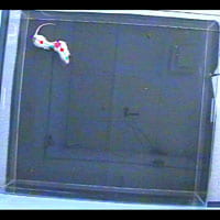
Social Interaction
Early life stress causes changes in social behavior. We use an open field task where experimental and control rats are matched with a conspecific rat. Tracked behaviors include locomotion, average distance apart from the conspecific, approach towards the conspecific, nose-to-nose contacts, and latency to first contact. We collect blood and brain tissue from the same rats who have been tested in the social interaction paradigm allowing us to correlate behavioral scores to biomarker levels.
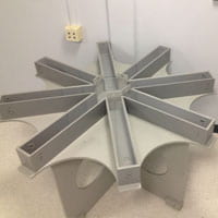
Win Shift Maze
The win-shift maze is a cognitive task that tests short and long term memory of Sprague Dawley rats. The paradigm allows us to compare a prefrontal cortex dependent behavior on rats who have either been exposed to early life stress or not. We collect bled and brain tissue from the same rats who have been tested on the win-shift maze, allowing us to correlate behavioral scores to biomarker levels.
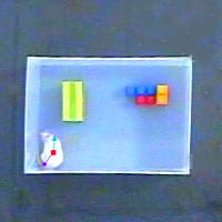
Novel Object Recognition
Novel object recognition is a minimally stressful task that evaluates memory alterations in rodents. The task assesses the ability of rodents to distinguish between a novel object and a familiar one within a habituated environment. The ability to recognize a familiar object is considered to be a hippocampus-PFC derived ability, rodents preferentially spend more time exploring the novel object, without any external cues.
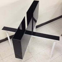
Elevated Plus/Zero Maze
Elevated Plus Maze (EPM) and Elevated Zero Maze (EON) are tests for assessing anxiety-like behavior in rodents. The EPM is a “+* shaped maze, consisting of two open and two closed arms, and the EOM is a circle, with two opposite areas enclosed and the other areas open. Both are elevated at 40 cm above ground. During the test, anxious rodents, due to their unconditioned fear towards open spaces, will be inclined to spend more time in closed spaces. Anxiety-like behavior is evaluated by measuring number of entries into either open or closed spaces.
Neural Tracing & Circuit Manipulation
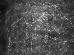
Basolateral Amygdala Prefrontal Cortex
Axons and axonal boutons in the prefrontal cortex (PFC) that originated in gluatamatergic cells in the basolateral amygdala (BLA). These axonal segments fluoresce due to a genetic tool called DREADDs (Designer Receptor Exclusively Activated by Designer Drugs). We inject the BLA’s of juvenile rats so that we can modulate the excitatory corticolimbic projections following early life adversity.
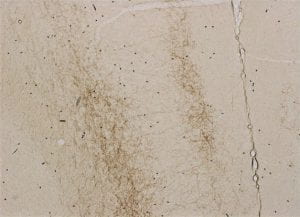
Neural Projections
We aim to understand the development of corticolimbic circuitry throughout early life and adolescence. One way to accomplish this is through anatomical tracing of neurons using mircoinjections of neuronal tracers into discrete areas of the brain. Following these microinjections we then measure the density of termianls in a projection site, like the prefrontal cortex. Shown above are neuronal projections from the basolateral amygdala in layers II and V of the prefrontal cortex.
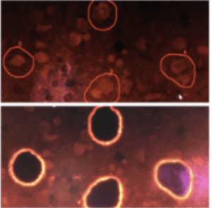
Laser Capture Microdissection
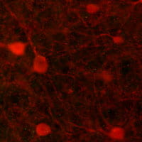
Laser Capture Microdissection (LCM) allows us to selectively investigate the properties of individual cell types (e.g., Parvalbumin+ cells, as seen here in red) within specified brain regions (e.g., PFC). This technique utilizes both Infrared and Ultraviolet lasers to ‘cut out’ and collect only the cells we are interested in studying. Once collected, mRNA can be extracted and analyzed using quantitative PCR. We carry out this research in conjunction with our collaborator, Dr. Greg Miller (Addiction Sciences Laboratory, NEU).
Immunofluorescence
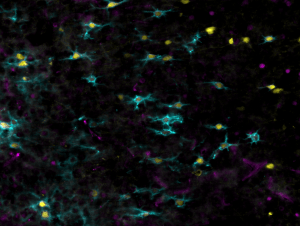
Perineuronal nets enwrapped in interneurons
Perineuronal nets (shown in blue) are components of the extracellular matrix that are important for synaptic stabilization in the brain. These structures preferentially enwrap the cell bodies of parvalbumin expressing interneurons (shown in yellow). Perineuronal nets are involved in regulating plasticity, as their emergence in development is related to the closure of childhood sensitive periods.
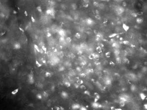
Interneurons (Parvalbumin)
Early life stress has been shown to affect the interneuron population in the prefrontal cortex. Parvalbumin (PVB) is a calcium binding protein located within interneurons in the brain. We use an antibody that binds to PVB in order to view Interneurons in the prefrontal cortex. We have completed stereological and exhaustive cell counts of PVB positive cells in the prefrontal cortex in order to see how early life stress impacts the interneuron population of the prefrontal cortex.
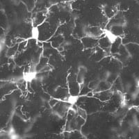
Microglia (IBA1)
IBA1 (ionizing calcium-binding adapter molecule 1) is calcium-binding protein that is specifically expressed by macrophages/microglial cells. Early life stress has been shown to cause increased microglial sensitization and expression within the PFC. We are interested in creating a developmental profile of microglial phenotype post maternal separation.
Assays
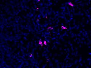
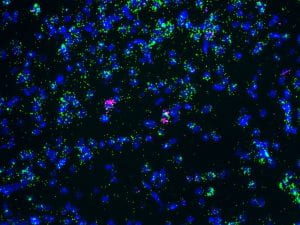
RNAscope Kiss 1 RNAscope POLR2A
Single-molecule visualization of RNA targets. Image shows Kiss1 (pink) and POLR2A (green) in the arcuate nucleus of the hypothalamus
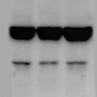
Western Blots
In our lab we use western blots in conjunction with co-immunoprecipitation in order to reveal if TATZA injections in the prefrontal cortex were successful in uncoupling the NR2A subunits of glutamate receptors in GABAergic interneurons of the prefrontal cortex. We have also used western blots to observe how various protein levels are affected by early life stress.
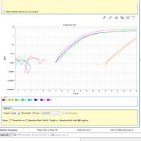
Quantitative PCR
We use quantitative PR to measure mRNA sequence expression that may be vital in the deregulation caused by early life stress. For example, we are using Taqman q-PCR to measure gene expression of cytokines such as TNFa and IL-1B, as well as the expression of microglia activation markers like MHCIL.
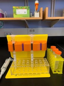
Microglia isolation involves dissociating rat brain tissue into a single cell suspension and magnetically labeling the CD11b+ cells. The cell suspension is passed through a column so that the unlabelled cells are filtered out and the CD11b+ labelled cells are collected. This technique can be followed by qPCR to measure gene expression in microglia specifically.